 A A
Acute Back Pain: Acute
low back pain, lower back pain or neck pain generally
lasts less than six months.
A few cases may resolve without medical attention, although
many reoccur.
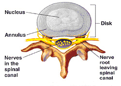
Annulus
Fibrosus (an-U-lus, fi-BRO-sus) The
thick fibrous outer ring of the intervertebral disc that
surrounds and encloses a gelatinous or jelly-like substance
(nucleus pulposus) in the center of the disc.
It provides support and stability to the disc.
B
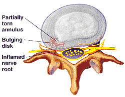
Bulging
Disc A broad-based
extension outward of the intervertebral disc in
one or all directions. In a bulging disc, the fibrous
outer ring (annulus fibrosus) of the disc and
vertical ligament in the back of the spinal vertebrae (posterior
longitudinal ligament) are both intact but can be
weakened. This often results from pressure on the disc
caused by activities such as lifting heavy objects and
straining the back or neck.
C
Chronic
Back Pain: Chronic low back
pain, lower back pain or neck pain generally persists
beyond six months.
If you are experiencing chronic back pain or chronic
neck pain, you IDD Therapy® may be an appropriate
treatment for you.
Coccyx: Two
to four tiny, partially fused vertebrae at the
end of the sacrum.

click image to enlarge
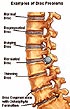
D Degenerative Disc Disease (Disc Degeneration) A
shrinking or narrowing of the disc space often accompanied
by bony spurs on the vertebra known as osteophytes.
Degeneration of the disc over time produces low-grade inflammation and
irritation and is a major cause of chronic low back
pain. Because the discs in the spine do not have
a dedicated blood supply, the discs must rely on
a process
called diffusion to receive their supply of water,
nutrients, and oxygen. If the flow of these elements
is disrupted, the vertebral discs can degenerate.
Disc degeneration can be mild, moderate or severe.
click image to enlarge
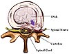
Disc
(Intervertebral Disc) The tough, elastic
structure that is between the bodies of spinal vertebrae.
The disc consists of an outer annulus fibrosus enclosing
an inner nucleus pulposus. The discs serve
as cushioning between and shock absorbers for the
spinal vertebrae.
E Extruded disc or Extrusion This
refers either to an leakage of part of the soft, gelatinous
central portion (nucleus pulposus) of the intervertebral
disc through a defect in the fibrous outer ring
(annulus fibrosus), a piece of the annulus
fibrosus that is torn away and hanging off of the
disc or both. This often results in pain that can be
localized or may extend into the leg and buttock causing sciatica.
F Facet or Facet Joint (fuh-SET) Each
spinal vertebra contains two upper (superior)
and two lower (inferior) facets along the back arches
of the bone. The facets are the sites where the adjacent
vertebra above and below connect with a single vertebra.
This connection forms the facet joint that allows
motion in the spinal column.
 click
images to enlarge click
images to enlarge 
Facet
Syndrome This is a condition where there
is degeneration and/or inflammation of the
facet joint leading to pain and some subsequent immobility
at the specific facet joint or spinal vertebral region.

click image to enlarge
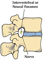
Foramen or Foramina A
natural opening or passage in bone. In the spine,
these are termed neural foramina or neuroforamina.
They are canals formed by the union of two adjacent
facets. These canals allow for the passage of the spinal
nerve root and spinal nerve as it travels from
the spinal cord to the rest of the body.
H
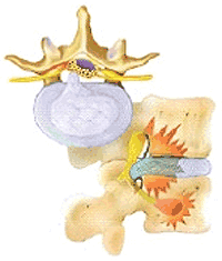
Herniated Disc The local protrusion
of disc material resulting from a tear in the outer
fibrous ring (annulus fibrosus) of the disc.
This allows for part or all of the soft, gelatinous
inner portion (nucleus pulposus) of an intervertebral
disc to escape from its center causing a flattening
of the disc (loss of disc height), decreased
shock absorbing and cushioning function and a chemical
irritation of the spinal nerve root resulting
in pain. This pain can be localized or may extend
into the leg and buttock causing sciatica.
Pain from a herniated neck disc may spread into
the shoulders or arms. Extension of pain into the
arms
or legs may be associated with radiculitis or
radiculopathy.
 click
image to enlarge click
image to enlarge 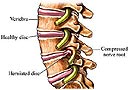
Herniated Disc in the Cervical (Neck)
Spine There are seven vertebrae in
the cervical spine (neck), referred to as C1 to C7.
The statistics regarding herniated disc in the
neck are approximately: C6-7 (69%), C5-6 (19%), C7-T1
(10%) and C4-5 (2%).
Herniated Disc in the Lumbar
(Low Back) Spine There are five vertebrae in
the lumbar spine (lower back), referred to as L1
to L5. The statistics regarding herniated disc in
the low back are approximately: L4-5 (35%), L5-S1
(27%), L3-4 (19%), L2-3 (14%) and L1-2 (5%).
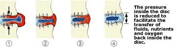
I IDD Therapy® IDD
Therapy® or Intervertebral Differential Dynamics
Therapy® is a proven non-surgical, non-invasive
treatment for the relief of lower back pain and neck
pain. It is an innovative approach effective in treating
herniated discs, bulging discs, degenerative disc disease,
posterior facet syndrome, sciatica and acute or chronic
neck and back pain. It is a safe, painless and comfortable
treatment found to be 86% to 92% successful in relieving
lower back pain and neck pain symptoms.
Inflammation The
result of localized injury or damage, which starts
a cascade of responses leading to swelling (edema),
warmth, red color (erythema) and pain in the area.
Inflammation triggers the pain receptors (nociceptors)
leading to both acute pain and chronic pain.
Intervertebral Canal A passageway
for the spinal nerve root formed by the joining of
two adjacent vertebrae at their facet joints. This
allows nerve fibers from the spinal cord to reach
the rest of the body. It is also called an intervertebral
foramen, neural foramen or neuroforamen.
Intervertebral Disc See Disc. Intravertebral Foramen A
canal formed by the bony arches on the back of the vertabrae
through which passes the spinal cord. The intravertebral
foramen of all the vertebrae form the length of the spinal
canal.

L Ligament A band
of flexible, fibrous connective tissue that
is attached at the end of a bone
near a joint. The main function of a ligament is to
attach bones to one another, to provide stability
of a joint,
and to prevent or limit some joint motion.
Lordosis This
is the normal backward curvature of the spine in the
region of the neck and low back. An increase or decrease
in the natural lordotic curve of the lumbar (low back)
or cervical (neck) spines is often abnormal.
Lumbago
or Lumbalgia Terms used to specify pain
in the lumbar region of the spine. They are synonymous
with
low back pain or lower back pain.
Lumbar The
lower part of the spine, located between the thoracic
spine (midback) and the sacrum (tailbone). The lumbar
spine consists of five vertebrae, named
in sequence from top to bottom as L1 through
L5. The
lumbar spine
supports most of the body's weight and absorbs
large amounts of stress.
N Nerve Root An
extension of the nervous system from the spinal
cord, within the spinal canal, to the rest
of the body. There
are two spinal nerve roots and spinal nerves
that exit from the sides of the spinal cord
through the vertebral
neural foramina at each vertebral level. These
nerves travel on to innervate the arms, legs
and remaining
body structures.
Nucleus Pulposus (NOO-klee-us,
puhl-PO-sus) The
semigelatinous tissue in the center of
an intervertebral disc. It is surrounded by
and contained within the annulus
fibrosus, which prevents this material
from protruding outside the disc space.
O Osteophytes Bony spurs
or outgrowths of the bone resulting from wear and tear.
These bony growths may or may not irritate the surrounding
tissue or nerves causing inflammation and pain.
Osteophytes and osteophytic growth are generally associated
with Osteoarthritis.
P Piriformis
Syndrome – A
group of symptoms that begin as a result of
spasm in the piriformis
muscle, which compresses or irritates the sciatic
nerve causing pain down the leg, usually as far
as the knee. The piriformis muscle is located
in the gluteal region and helps rotate the hip
out to the side. The sciatic nerve runs beneath
the piriformis muscle as it exits the pelvis
to enter the leg. This syndrome is treatable
with osteopathic manipulation and stretching
exercises.

click image to enlarge
R Radiculitis Inflammation
of a spinal nerve root in the area between the spinal
cord and the intervertebral canal. This is usually
associated with a bulging or herniated disc.
Radiculopathy Disease
or compression of the nerve root leading to serious
neurological problems with symptoms such as pain,
numbness or tingling
in a leg or legs.
Ruptured Disc See Herniated
Disc.
S Sacrum The
sacrum is a triangular bone consisting
of five fused vertebrae that
have no intervertebral discs. Its forms
the base of the spinal column and the keystone
of the pelvis. It is also known as the tailbone.
The sacrum
is joined
above with the lumbar spine at L5 forming the lumbosacral
junction and the two hip bones (ilia) on either
side forming the left and right sacroiliac joints.

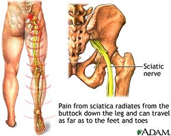
Sciatica Sciatica
refers to a pain felt along the length
of the sciatic nerve. The pain is usually
felt in the buttock and spreads down
the back of the leg to below the knee
and sometimes down to the foot. Sciatica
is one of the most common forms of pain
caused by compression of the spinal nerves.
The leg pain is often much worse than
the back pain. It is estimated that up
to 40% of people experience pain caused
by compression of this nerve at some
point in their lifetime. The six most
common causes of sciatic nerve compression
are: (1) a bulging disc or herniated
disc (2) lumbar spinal stenosis (3) spondylolisthesis
(4) trauma (5) piriformis syndrome, and
(6) spinal tumors.
Sciatic Nerve The sciatic
nerve is the largest and longest
nerve in the body. It originates from
five separate spinal nerves (L4-S3),
which join together to form a single
nerve called the sciatic nerve.
This nerve passes through the sciatic
foramen in the gluteal area to the
rest of the leg. In the leg, the nerve
separates in to a number of branches.
The nerve and its nerve branches enable
movement (motor function) and feeling
(sensory function) in the entire leg
and foot. The sciatic nerve may
sometimes be compressed by the piriformis
muscle as it exits the pelvis to enter
the leg.
Scoliosis Sideways
(lateral) curvature of the spine.
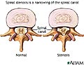
Spinal Stenosis Narrowing
of a space in the spine that part of
the nervous system travels through. This
results in compression of the nerve roots
in the neural foramen or spinal cord
in the spinal canal by bony spurs, a
bulging disc or a herniated disc. This
occurs most often in the lumbar spine
(low back) but also occurs in the cervical
spine (neck) and less often in the thoracic
spine (middle back). Spinal stenosis
is most often caused by degeneration
of the discs between the vertebrae due
to osteoarthritis.
 click
images to enlarge click
images to enlarge 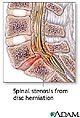
Spinal Disc See Disc
(Intervertebral).
Spinal Column See Spine.
Spinal Canal The
long bony channel formed by the intravertebral
foramen of all the vertebrae in order
to house and protect the spinal cord
and nerve roots.
Spinal Cord The
spinal cord is the central trunk of nerves
that connects the brain with the rest
of the body. It is enclosed in the spinal
canal. Each nerve root passes from the
spinal cord to the body through small
openings (foramen) bounded on one side
by the disc and the other by the facets.
When the spinal cord reaches the lumbar
region, it splits into four bundled strands
of nerve roots called the cauda equina
(meaning horsetail in Latin).
Spinal Fusion A
surgical procedure to permanently connect
two or more vertebrae in order to prevent
motion.
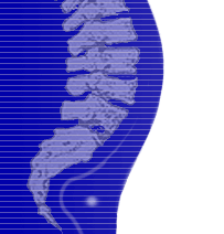
Spine The
flexible bony column extending from the
base of the skull to the tailbone. The
spine is the central support structure
of the body's bony framework. It is made
up of 33 bones, known as vertebrae. The
first 24 vertebrae are separated by intervertebral
discs and are bound together by ligaments
and muscles. Five vertebrae are fused
together to form the sacrum and 4 vertebrae
are fused together to form the coccyx.
There are four regions of the spine,
the cervical spine or neck, thoracic
spine or midback, lumbar spine or low
back and the base of the spine in the
pelvic area. The spine is also referred
to as the vertebral column, spinal column,
or backbone.
Spondylitis Inflammation
of the vertebrae.
Spondylolisthesis The
forward displacement of one vertebra
on another caused by a defect in the
bone between the superior and inferior
facets. The vertebra with the defect
and the spine above that vertebra are
slipped forward in relationship to the
vertebrae below. This condition can vary
in degree of displacement. It is usually
due to a developmental defect or the
result of a break in one or both of the
bony arches in back of the vertebra.

click image to enlarge
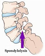
Spondylolysis Displacement
of one vertebra over another associated
with a break in the back arch of the
vertebra. This results from a defect
in the arch between the superior and
inferior facets of the vertebrae.
It may be one-sided or occur on both
sides and is usually due to a developmental
defect but may be due to a fracture.
Spondylosis Degenerative
changes of the vertebrae, vertebral joints, intervertebral
discs or the surrounding ligaments,
mainly due to osteoarthritis. This leads
to stiffness and may cause pain, numbness
or tingling in the arms or legs.
V
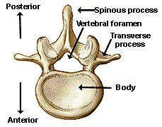
Vertebra One
of the 33 bones of the spinal column.
A cervical, thoracic, or lumbar vertebra
has a circular body in front and two
arches behind (composed primarily of
the laminae and pedicles as well as the
other structures). Together these structures
form a canal that houses and protects
the spinal cord as it descends from the
brain to the tailbone. The plural of
vertebra is vertebrae.
|